Methods in Molecular Biology 773: 319-327 (2011)
In vivo 1H-NMR microimaging during seed imbibition, germination, and early growth
University of Freiburg, Faculty of Biology, Institute for Biology II, Botany / Plant Physiology, Schänzlestr. 1, D-79104 Freiburg, Germany, Web: 'The Seed Biology Place' http://www.seedbiology.eu (G. L.-M.)
Department of Biological Sciences, Simon Fraser University, 9 8888, University Drive, Burnaby BC, V5A 1S6, Canada (K. M., A. K.)
Steacie Institute for Molecular Sciences, National Research Council Canada, Ottawa, ON, Canada (V. T.)
Chapter 18 in: Allison R. Kermode (ed.), Seed Dormancy: Methods and Protocols
DOI 10.1007/978-1-61779-231-1_18, © Springer Science+Business Media, LLC 2011
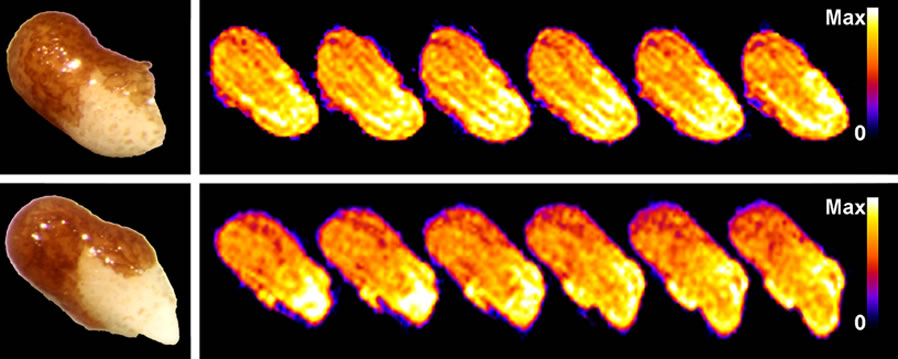
Fig. 1. Noninvasive in vivo 1H-NMR microimaging of water uptake and distribution during tobacco seed germination.
The spatial distribution of protons within the seed tissues is visualized with false colors as shown. The NMR microimages were obtained with 30 mm spatial resolution.
Also shown are corresponding microphotographs of seeds in the testa rupture (top left) and endosperm rupture (bottom left) stages.
Modified from Manz et al. (2005), http://www.plantphysiol.org, Copyright American Society of Plant Biologists.
| Article in PDF format (0.3 MB) |
|
|
|
The Seed Biology Place |
Webdesign Gerhard Leubner 2000 |