Plant Physiology 160: 1551-1566 (2012)
Distinct cell wall architectures in seed endosperms in representatives of the Brassicaceae and Solanaceae [W][OA]
Centre for Plant Sciences, Faculty of Biological Sciences, University of Leeds, Leeds LS2 9JT, UK (KL, PK)
Wageningen Seed Lab, Laboratory of Plant Physiology, Wageningen University, Droevendaalsesteeg 1, 6708 PB, Wageningen, The Netherlands (BD, LB)
Department of Molecular Plant Physiology, Utrecht University, 3584 CH Utrecht, The Netherlands (BD, LB)
University of Freiburg, Faculty of Biology, Institute for Biology II, Botany/Plant Physiology, D-79104 Freiburg, Germany (TS*, GLM*)
ARC Centre of Excellence in Plant Cell Walls, School of Botany, University of Melbourne, Parkville, Victoria 3010, Australia (BD, LB)
* Current Address: School of Biological Sciences, Royal Holloway, University of London, Bourne Building 3-30, Egham, Surrey, TW20 0EX, UK
Received July 13, 2012; Accepted September 4, 2012; Published September 6, 2012.
DOI:10.1104/pp.112.203661
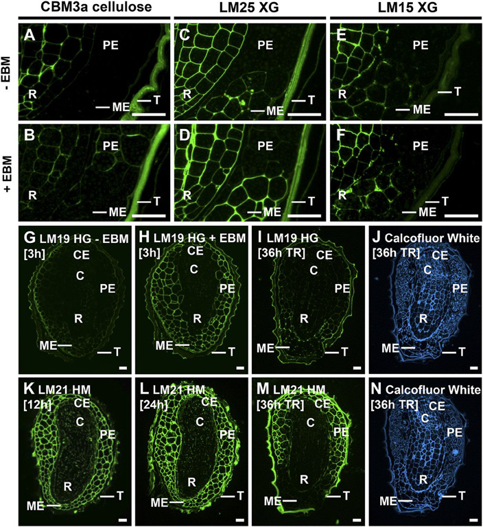
Figure 6. Medial longitudinal sections of tobacco seeds with enzymatic deconstruction and during germination. Three-hour-imbibed mature tobacco seed sections were treated with EBM or a buffer control prior to immunolabeling.
A and B, CBM3a cellulose was unmasked in PE and CE cell walls following EBM treatment.
C and D, LM25 XG epitopes were unmasked in ME following EBM treatment.
E and F, LM15 XG epitopes were not substantially unmasked in ME following EBM treatment.
G and F, LM19 HG was weakly detectable in the endosperm of 3-h-imbibed seeds and was unmasked in all endosperm cell walls by EBM treatment.
I, The LM19 epitope was unmasked in the ME at testa rupture (TR) at 36 h.
K to M, Immunolabeling of germinating tobacco seeds with LM21 HM revealed the specific degradation of heteromannan (HM) at the ME at testa rupture. R, Radicle; C, cotyledons; T, testa. Bars = 50 mm.
| Article in PDF format (1.5 MB) Supplementary data file (2 MB) |
|
|
|
The Seed Biology Place |
Webdesign Gerhard Leubner 2000 |