Plant Physiology 150: 1855-1865 (2009)
In vivo cell wall loosening by hydroxyl radicals during cress (Lepidium sativum L.) seed germination and elongation growth [W][OA]
The Edingburgh Cell Wall Group, Institute of Molecular Plant Sciences, The University of Edinburgh, The King's Buildings, Mayfield Road, Edinburgh EH9 3JH, United Kingdom (R.A.M.V., S.C.F.)
Commissariat à l'Energie Atomique (CEA), iBiTecS, CNRS URA 2096, Service de Bioénergétique, Biologie Structurale et Mécanisme, 91191 Gif-sur-Yvette Cedex, France (A.K.-L.)
Received April 9, 2009; accepted May 29, 2009; published June 3, 2009
www.plantphysiol.org/cgi/doi/10.1104/pp.109.139204
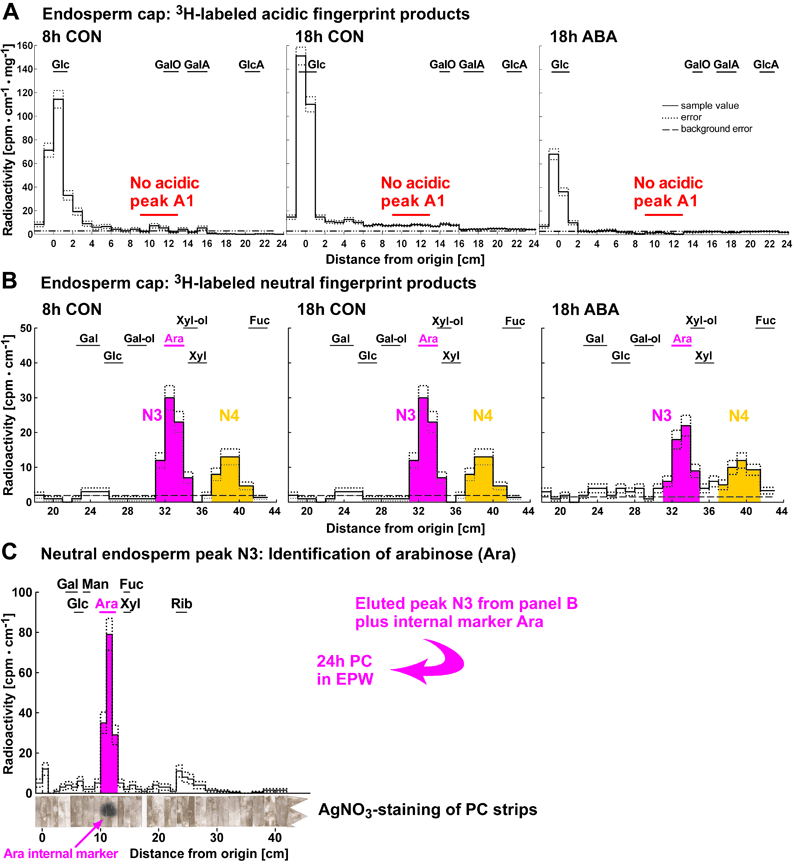
Figure 4. Detection of in vivo •OH attack on cress seed endosperm cap cell walls by 3H-fingerprinting and identification of the neutral compound observed as peak N3 as arabinose. 3H-fingerprints of cap samples (A and B) differ quantitatively and qualitatively from those of radicle samples (see Fig. 3). (A) Representative electrophoretic 3H-fingerprints of 3H-labeled products from cap samples. Signal intensity in the scintillation count is plotted against distance from the origin after high-voltage paper electrophoresis (PE) at pH 3.5. No acidic peak was detected. (B) Representative chromatographic 3H-fingerprints of 3H-labeled products from cap samples. The samples were eluted from the fraction that comigrated with glucose during PE at pH 3.5 and re-run by paper chromatography (PC). (B and C) The neutral compound observed as peak N3 was identified as arabinose (Ara) by PC in different solvents with reference to internal and external markers (Fry, 2000). Peak N4 remains unidentified. Peak N3 comigrates with an external arabinose standard in butanol:acetic acid:water (12:3:5, v/v, ‘BAW’). (C) Peak N3 was eluted and re-run by PC in ethyl acetate:pyridine:water (8:2:1, v/v, ‘EPW’). Again, the peak comigrates with the external marker arabinose. The internal marker arabinose (arrow) comigrated with the EPW peak, as shown by AgNO3-staining of the strips of chromatography paper after recovery from the scintillation fluid. For abbreviations of acidic and neutral markers see Fig. 3.
| Article in PDF format (1.7 MB) |
|
|
|
The Seed Biology Place |
Webdesign Gerhard Leubner 2000 |